Mapping Gene Expression in the Mouse Lungs from Images to Scaffold
Overview
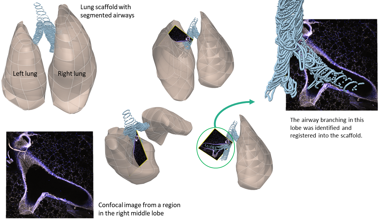
A 3D scaffold of the mouse thoracic cavity created using segmentation of longitudinal microCT scans from the SIMBA VIA (Vision and Image Analysis) public database is visualized. The trachea and main bronchi are also shown as rings of segmented points. Confocal stained images from Taylor-Clark group for mouse lungs are embedded into the scaffold. The confocal images were obtained from the right middle lobe (RML). The scaffold was registered to ensure alignment with the airway branch visible on the confocal. This use-case is one of many that link an organ systems with the brainstem.
Step-by-step instructions
Follow these step-by-step instructions to familiarise yourself with the flow of the web interface.
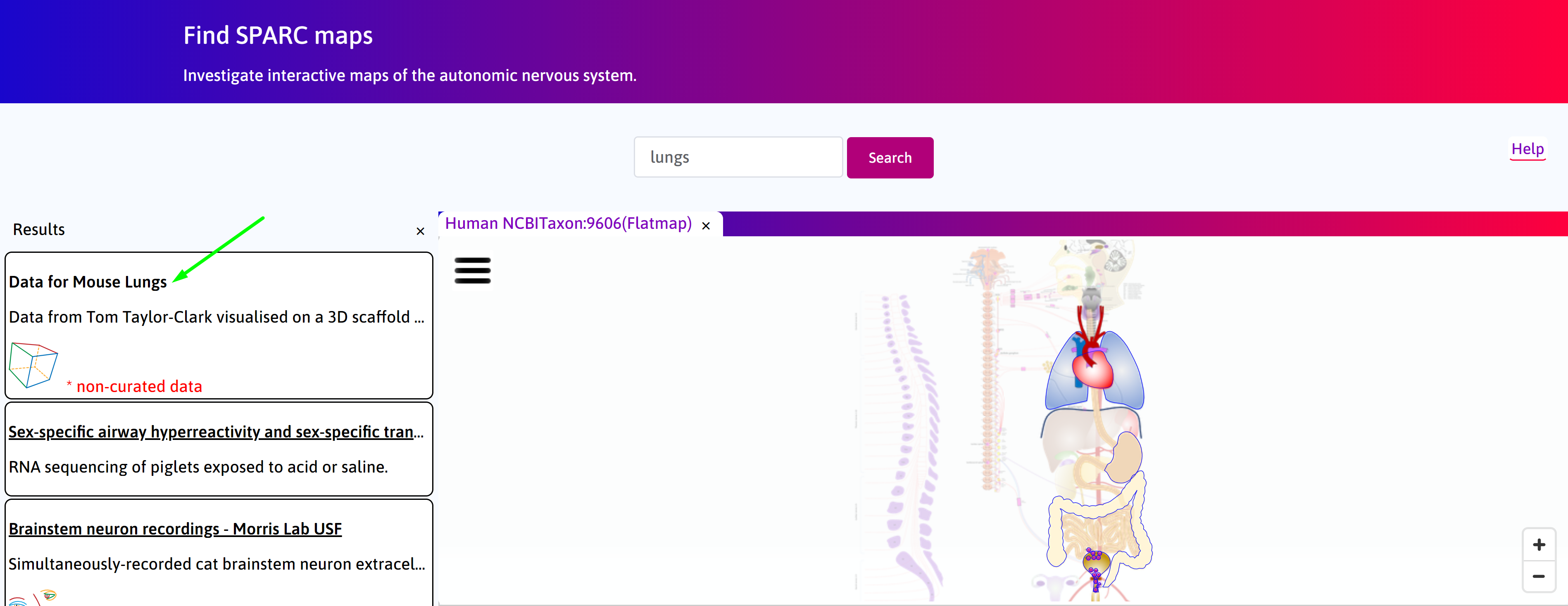
Step 1. The default Result column displays the use cases available on the portal. Click on the
Mapping Gene Expression in the Mouse Lungs from Images to Scaffold box, then on ![]() icon.
icon.
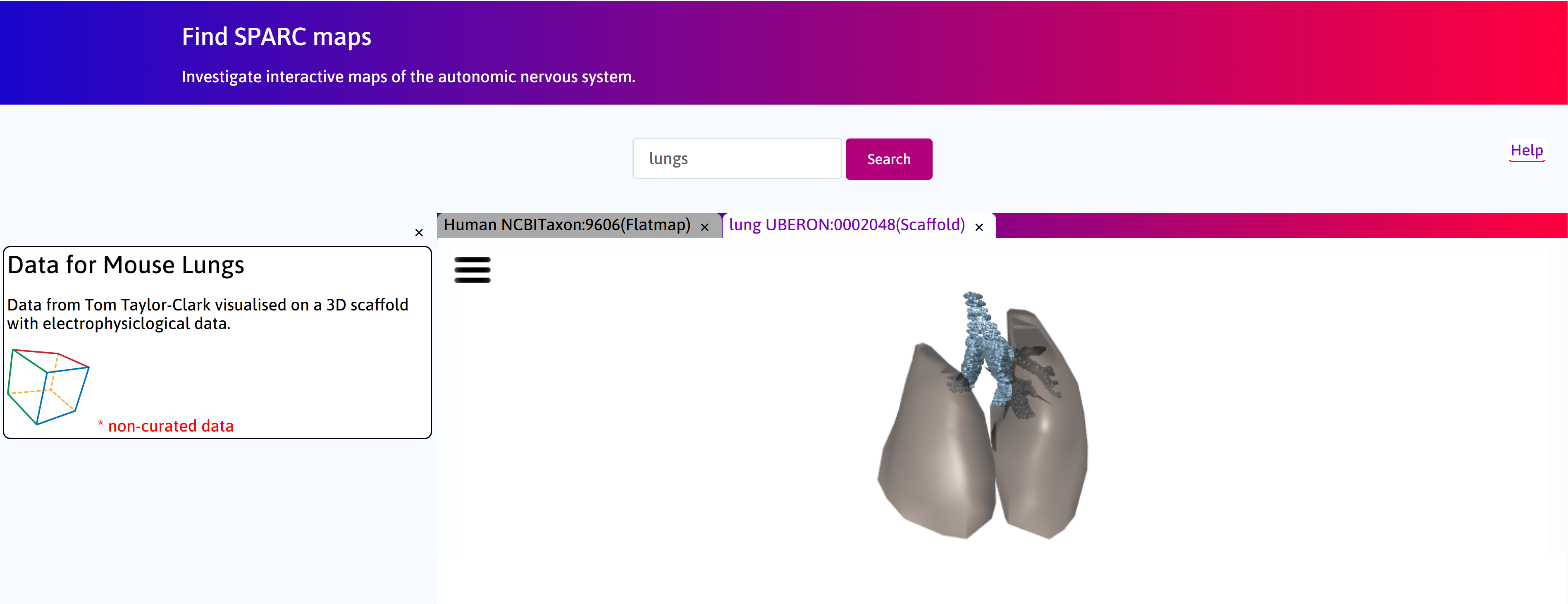
Step 2. Allow model to load.
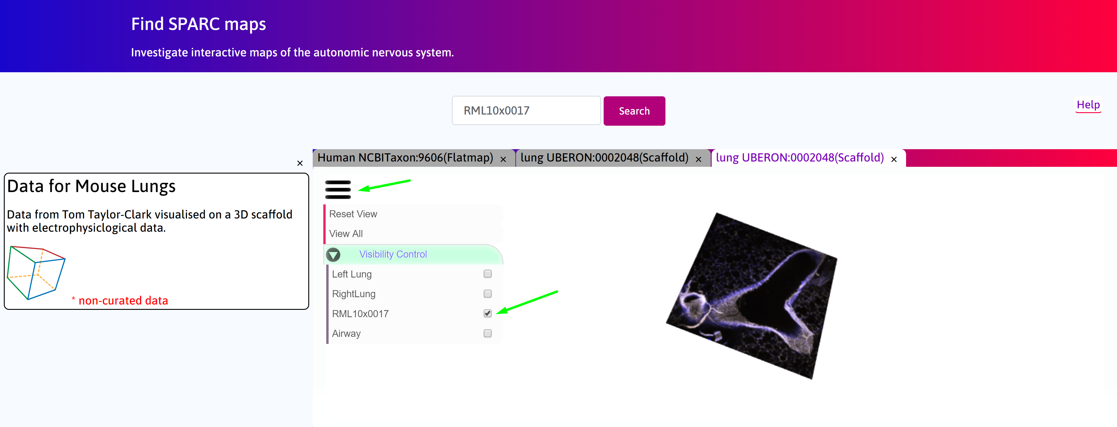
Step 3. To view only the confocal image in the right middle lobe (RML), select only RML10x007 checkbox after clicking on  icon for drop-down menu.
Scaffold Generation
A diagram and video are below, detailing the workflow for the generation of an anatomically-based 3D thoracic shape of the lungs.