Mapping the Mouse Heart Neurites from Image to Scaffold
Overview
Immunohistochemical mapping of neural pathways in cleared heart (sham heart 4) stained with PGP9.5 (glycoprotein surface axonal antibody labelling) from the Shivkumar/Pradeep group is displayed in a 3D mouse heart scaffold that has been fitted to the segmented heart surface data from Sham Heart 44, which provides a more extensive geometric dataset. Future experiments are likely to provide much better quality mouse data for both the heart’s anatomy and its embedded neurons.
Step-by-step instructions
Follow these step-by-step instructions to familiarise yourself with the flow of the web interface.
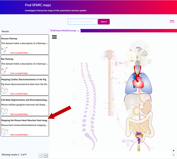
Step 1. The default Result column displays the use cases available on the portal. Click on the Mapping the Mouse Heart Neurites from Image to Scaffold box.
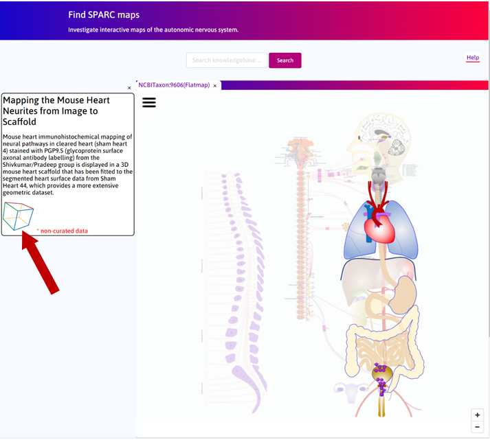
Step 2 Click on the ![]() to open the Scaffold Viewer tab to visualise the heart scaffold.
to open the Scaffold Viewer tab to visualise the heart scaffold.
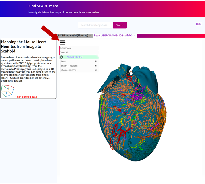
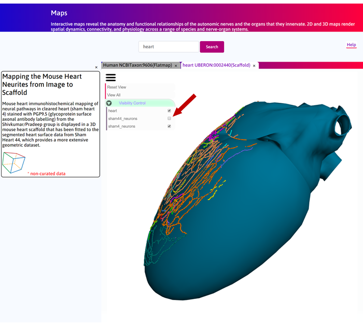
Step 3. In the Scaffold Viewer tab, click on  to open the control panel. There are three objects visible: 1) fitted heart scaffold, 2) sham heart 44
neurite tracing, 3) sham heart 4 neurite tracing.
to open the control panel. There are three objects visible: 1) fitted heart scaffold, 2) sham heart 44
neurite tracing, 3) sham heart 4 neurite tracing.
Step 4. Turn off Sham 4 neurons to only visualise the scaffold and Sham 44 neurons.
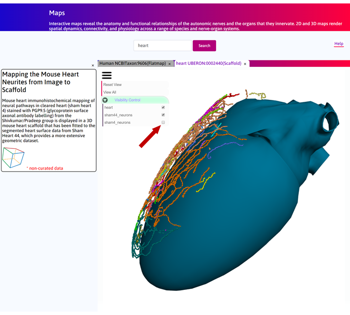
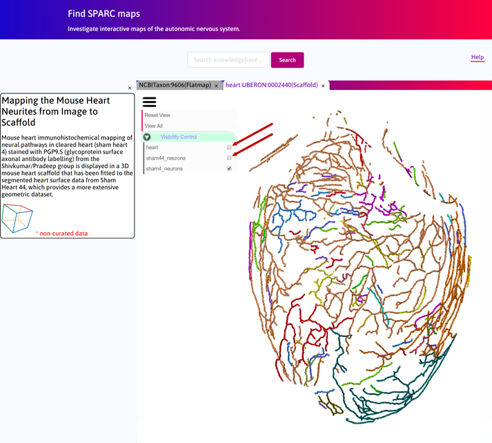
Step 5. Turn Sham 4 neurons back on and now turn off Sham 44 neurons to visualise the registered neurons on the scaffold surface.
Step 6. To visualise only the neurons, turn off the scaffold.
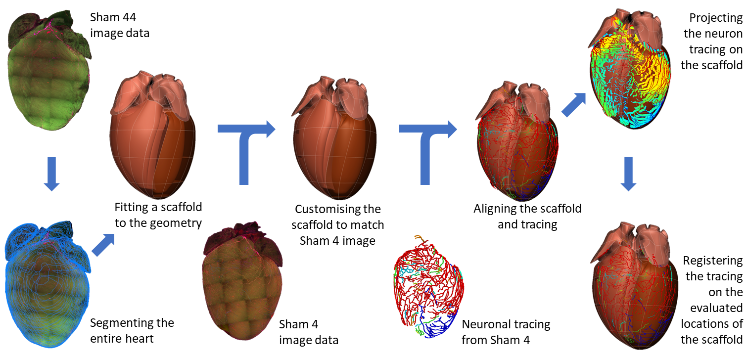
Scaffold Generation
The following figure illustrates an overview of the workflow for the generation of the 3D scaffold.
Below is an introductory video which explains the mapping of the mouse heart neurites from image to scaffold.